
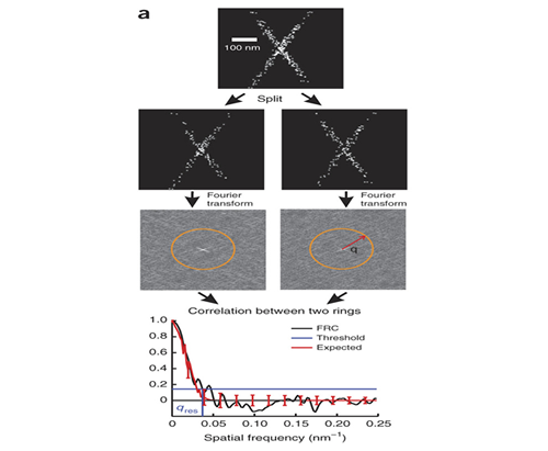
Single molecule localization based super-resolution techniques can provide images with resolution several times better than the diffraction limit. However, the achieved image resolution in a particular experiment is difficult to quantify because it cannot be given by specifications of the instrument and is instead a complicated function of the localization density, localization error, and the sample itself. Together with the Quantitative Imaging group at Delft (PI Bernd Rieger) we developed a method for using Fourier Ring Correlation to quantify image resolution for these methods.
R. P. J. Nieuwenhuizen, K. A. Lidke, M. Bates, D. L. Puig, D. Grünwald, S. Stallinga, and B. Rieger. (2013) "Measuring image resolution in optical nanoscopy.," Nature methods 10, 557–562 .
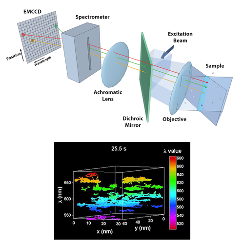
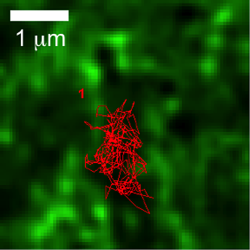
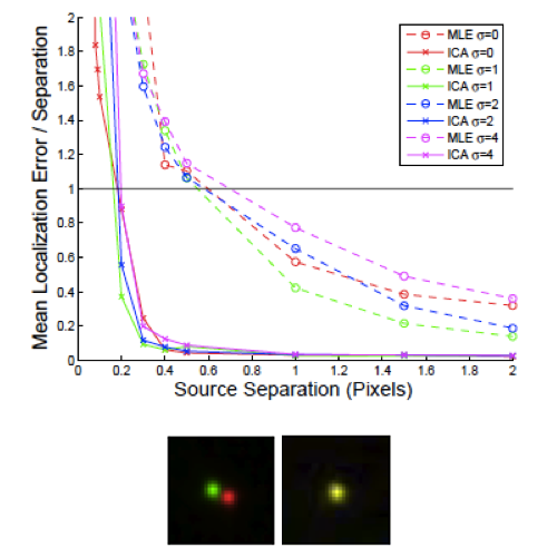
We have developed a novel high-speed hyperspectral microscope (HSM) to perform single particle tracking of up to 8 spectrally distinct species of quantum dots (QDs) at 30 frames per second. The distinct emission spectra of the quantum dots allows localization with ~ 10 nm precision even when the probes are clustered at spatial scales below the diffraction limit. The HSM is now being used to study the dynamics and stoichiometry of several types of membrane signaling complexes.
Cutler PJ, Malik MD, Liu S, Byars JM, Lidke DS, et al. (2013) Multi-Color Quantum Dot Tracking Using a High-Speed Hyperspectral Line-Scanning Microscope. PLoS ONE 8(5): e64320. doi:10.1371/journal.pone.0064320
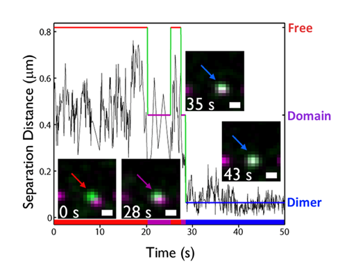
Ligand-induced signaling by the epidermal growth factor receptor (EGFR or HER1 or erbB1) drives cell growth and survival, with roles in normal development and disease pathogenesis. A wealth of structural knowledge supports a model of signal initiation through the formation of back-to-back erbB1 dimers. However, conclu¬sions about the size and ligand-occupancy of the erbB1 signaling complex remain controversial. We used two-color single quantum dot tracking to directly observe and quantify erbB1 homodimerization on living cells. To quantify the kinetics of dimerization, a 3-state Hidden Markov Model was developed to extract transition rates between free, co-confined, and dimer states. It was found that 2 ligand-bound receptors form more stable dimers than resting receptors, linking ligand occupancy to dimer stability. Furthermore, actin-based confinement was found to promote receptor dimerization.
S.T. Low-Nam, K.A. Lidke, P.J. Cutler, R.C. Roovers, P.M.P. van Bergen en Henegouwen, B.S. Wilson, D.S. Lidke. (2011) "ErbB1 dimerization is promoted by domain co-confinement and stabilized by ligand." Nature Structural & Molecular Biology. 18:1244-1249
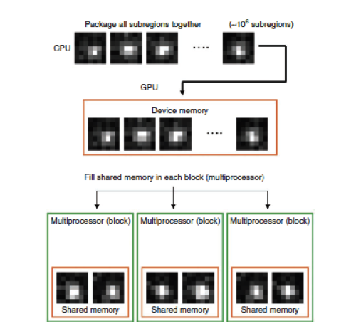
Modern Graphics Processing Units (GPUs) have approximately two orders of magnitude higher floating point performance than that of CPUs. This is largely due to the massively parallel architecture of such cards - GPUs have hundreds to thousands of processing cores. My lab has developed efficient routines for estimation of fluorophore positions that run on GPU architecture using NVIDIA's CUDA interface. Our original work, (Smith, Nature Methods, 2010) was the first to use GPUs for this task and demonstrated a maximum likelihood estimation method that could process 105 particle localizations per second. Implementations on newer GPUs now achieve over 106 fits per second. This approach took the most demanding task of super-resolution analysis and made it trivial, removing the need for less accurate approximations in order to achieve reasonable speed.
Smith CS, Joseph N, Rieger B, Lidke KA. (2010) "Fast, single-molecule localization that achieves theoretically minimum uncertainty." Nat Methods. 2010;7(5) p. 373-5.
The concept of actin "confinement zones" has been proposed for more than a decade. Through simultaneous observations of quantum dot (QD)-labeled FcεRI motion and GFP-tagged actin dynamics, we provided the first direct evidence for actin barriers that influence protein diffusion on the time scale of seconds and length scale of microns. Dynamic reorganization of actin structures occurs over seconds, making the location and dimensions of actin-defined domains time dependent. We also designed a novel assay to quantify receptor immobilization kinetics in response to cross-linking by multivalent antigen. This assay revealed that dramatic changes in receptor mobility occur within seconds of antigen binding and the kinetics are dependent upon an intact actin cytoskeleton. These new insights not only confirm that actin acts as a diffusion barrier, but also demonstrate a significant role for actin in modulating protein-protein interactions and ligand binding response.
N.L. Andrews, K.A. Lidke, J.R. Pfeiffer, A.R. Burns, B.S. Wilson, J.M. Oliver and D.S. Lidke. (2008) "Actin restricts FcεRI diffusion and facilitates antigen induced immobilization." Nature Cell Biology, 10:955-963
In 2005, we were the first to propose and demonstrate that the blinking of fluorophores could be used to identify individual emitters and thereby allow localization with precision better than the diffraction limit and better than direct fitting to the temporal cumulative image. We proposed that high resolution images of structures could be generated from the localization of many fluorophores and named the method 'Pointillism'. Our paper used Quantum Dots, which are bright and photo-stable but have a high minimum duty cycle that limited the labeling density. We proposed that other methods of generating blinking or dark states could be used including triplet state blinking and photo-chromic fluorophores. These methods have since been used for fluorescence superresolution.
Lidke KA, Rieger B, Jovin TM, Heintzmann R. (2005) "Superresolution by localization of quantum dots using blinking statistics." Opt Express. 2005;13(18) p. 7052-62.